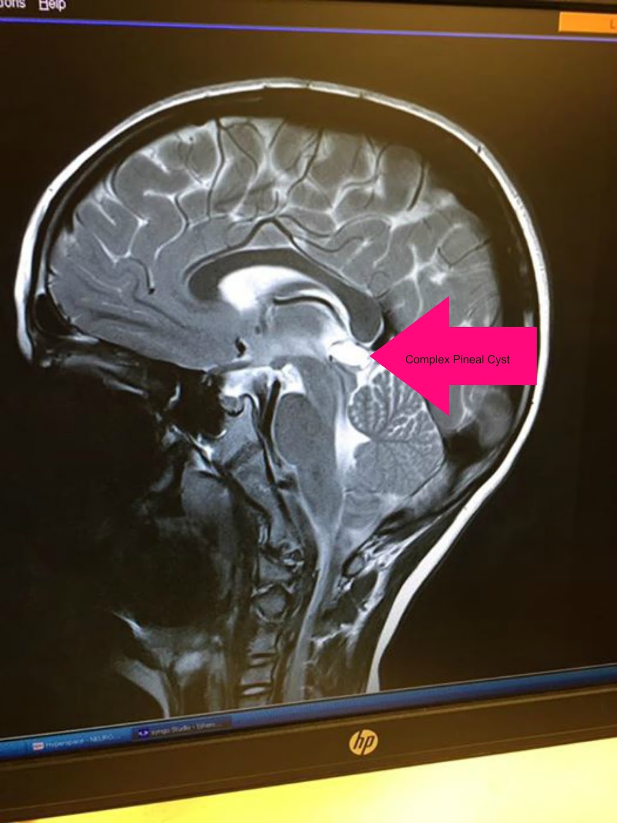
11++ Pineal Gland Cyst Mri Image
Pineal Gland Cyst Mri Image. It has been our experience, however, that when fully and completely imaged—which necessitates using postcontrast mr imaging—most of these tumors bear little resemblance to typical pineal cysts. Pineal cysts (pc) are cysts which are frequently detected incidentally in brain magnetic resonance imaging (mri).

Found a frequency of 23% in brain scans (with a mean. My pituitary was being imaged for thyroid problems i've been experiencing for the past three months (fluctuating tsh leaning more toward hypo yet elevated rai uptake, a litany of symptoms like my neck being swollen and tight on one side, etc.). I believe mri is the best imaging modality for viewing the structure of parts of the brain.
piscine ronde intex pince a oeillets mode demploi perforateur sds max peinture abris de jardin bois
Pineal gland apoplexy mimicking as migrainelike headache
I saw the neurosurgeon on monday for the pineal cyst that was found on my mri last month. The finding that pineal cyst development, small changes in cyst size, and cyst involution can all be seen on serial mr imaging without the development of specific symptoms supports the suggestion that typical pineal cysts found incidentally on mr imaging may be followed on a clinical basis alone, rather than by imaging. The pineal gland is a small organ in the middle of the brain. Pineal cysts (pcs) are a benign lesion of the pineal gland that have been known to the medical community for a long time.

A 2007 study by pu et al. This patient had an incidental pineal cyst on mri done for pituitary adenoma. The natural course of pc morphology has not been well described. A pineal cyst seldom causes problems. These cysts are benign, which means not malignant or cancerous.

As a general rule, pineal cysts are described as such when they reach a size of at least 5 mm, but there are reports of pineal cysts less than 2 mm in diameter. I've posted about this before but it's been a little while. A pineal cyst usually only shows up on an imaging scan done for another reason. A.

Neurosurgeon says he doesn't think it's causing any of my issues and just keep treating for my lyme and recheck it in 6 months. Pineal cysts (pc) are cysts which are frequently detected incidentally in brain magnetic resonance imaging (mri). A pineal cyst seldom causes problems. I believe mri is the best imaging modality for viewing the structure of parts.

If a pineal cyst grows large, it may affect your vision. A pineal cyst seldom causes problems. A 2007 study by pu et al. With a prevalence rate of approximately 1% in the general population, pc is often a reason for medical counseling. The vast majority of cysts are simple, unilocular, and follow csf density (ct) and signal intensity (mri),.

Found a frequency of 23% in brain scans (with a mean. An mri should be able to visualize the pineal gland and determine whether it is solid, cystic or enlarged. Pineal cysts (pc) are cysts which are frequently detected incidentally in brain magnetic resonance imaging (mri). A pineal cyst usually only shows up on an imaging scan done for another.

Although pineal cysts are found with a frequency of over one third in autopsy series, prevalences reported in standard magnetic resonance imaging (mri) studies only range between 0.14% and 4.9%. In series of magnetic resonance imaging (mri) studies, the prevalence of pineal cysts ranged between 1.3% and 4.3% of patients examined for various neurologic reasons and up to 10.8% of.

With a prevalence rate of approximately 1% in the general population, pc is often a reason for medical counseling. With the intro duction of gadopentetate dimeglumine, we have noticed that these pineal cysts may enhance and resemble solid tumors on delayed images obtained after administration of the contrast medium. If the cyst reaches 15 mm or larger, however, it may.

This is usually because the cyst has grown so much that it has pressed into other parts of the brain. With a prevalence rate of approximately 1% in the general population, pc is often a reason for medical counseling. Such a growth can cause symptoms ranging in severity from nausea and headaches to coma. My pituitary was being imaged for.